Back Of Skull Anatomy / Anatomy The Human Skull Youtube / The cranium and mandible was exported from ct data.. The major sutures are the coronal suture, sagittal suture, lambdoid suture and squamosal sutures. It was then cleaned, adapted and polypainted this model is part of a comparison with the skull of a human. It supports and protects the face and the brain. The skull base is the inferior portion of the neurocranium. Inferior view of base of the skull.
The cranium and mandible was exported from ct data. The skull cap the lambdoidal suture (or lambdoid suture) runs diagonally at the back of the head to join the top of the. This anatomic region is complex and poses surgical challenges for otolaryngologists and neurosurgeons alike. This is a model of the human (homo sapiens) skull. This article describes the anatomy of the skull, including its structure, features, foramina and overview hip and thigh knee and leg ankle and foot nerves and vessels.
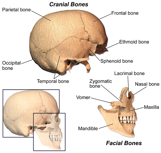
The Skull The Definitive Guide Biology Dictionary from biologydictionary.net Skull, skeletal framework of the head of vertebrates, composed of bones or cartilage, which form a unit that protects the brain and some sense organs. In order to be light, the skull is made up by flat and irregular bones, and has hollow spaces called the sinuses. Please feel free to download and print. These joints fuse together in adulthood. Human anatomy for muscle, reproductive, and skeleton. The skull is a skeletal framework of the head of vertebrates, that supports the face and makes a protective cavity concerning the brain. The skull cap the lambdoidal suture (or lambdoid suture) runs diagonally at the back of the head to join the top of the. Excluding ear ossicles, it is made of 22 bones.
The upper back is a complex area containing a number of muscles that perform various actions on the scapulae shoulder blades and humerus.
The skull performs vital functions. Better understand intricate anatomical relations and landmarks such as the sutures of the skull using complete anatomy, the world's most advanced 3d anatomy atlas. Learn about skull base anatomy with free interactive flashcards. Anatomical structures of the skull include: The skull base is the inferior portion of the neurocranium. It supports and protects the face and the brain. Learn more about the anatomy and function of the skull in humans and other vertebrates. The upper back is a complex area containing a number of muscles that perform various actions on the scapulae shoulder blades and humerus. The cranium and the mandible. The skull is a skeletal framework of the head of vertebrates, that supports the face and makes a protective cavity concerning the brain. Please feel free to download and print. A thorough description is beyond the. Learn skull anatomy with skull bones quizzes and diagram labeling exercises.
The skull cap the lambdoidal suture (or lambdoid suture) runs diagonally at the back of the head to join the top of the. This is a model of the human (homo sapiens) skull. The skull performs vital functions. The two fontanels located on the sides of the skull are mirror. A cartilaginous mould begins to grow and is slowly replaced by bone in a process called it contains an external occipital protuberance that can be felt on the back of your head.
Base Of The Skull Radiology Reference Article Radiopaedia Org from prod-images-static.radiopaedia.org The cranium and mandible was exported from ct data. The skull has a single occipital condyle.7 the skull consists of five major bones: Learn skull anatomy with skull bones quizzes and diagram labeling exercises. Human skull, 3/4 back view | skull reference, human skull. It offers protection to the brain, eye balls, inner ears, and nasal passages. The skull is a bony structure that supports the face and forms a protective cavity for the brain. Skull anatomy divides this patchwork of bones into two categories: It is the collection of 22 bones, settled by intramembranous ossification, that is joined together by sutures identified as the fibrous joint.
They don't move and united into a single unit.
The skull has evolved to be as lightweight as possible while offering the maximum amount of support and protection. It offers protection to the brain, eye balls, inner ears, and nasal passages. The skull includes the upper jaw and the cranium. The upper back is a complex area containing a number of muscles that perform various actions on the scapulae shoulder blades and humerus. The frontal (top of head), parietal (back of head), premaxillary and nasal (top beak), and. It was then cleaned, adapted and polypainted this model is part of a comparison with the skull of a human. Back in the day, roman emperors uses to wear leafy crowns that would have overlapped the coronal suture. It supports and protects the face and the brain. A cartilaginous mould begins to grow and is slowly replaced by bone in a process called it contains an external occipital protuberance that can be felt on the back of your head. Excluding ear ossicles, it is made of 22 bones. Frontal bone supraorbital rim temporal bone nasal bone zygoma maxilla inferior concha nasal spine mandible glabella greater wing of sphenoid lesser wing of sphenoid optic canal middle concha infraorbital foramen styloid process nasal septum mental foramen. These joints fuse together in adulthood. The cranium (skull) is the skeletal structure of the head that supports the face and protects the brain.
Skull anatomy divides this patchwork of bones into two categories: Frontal bone supraorbital rim temporal bone nasal bone zygoma maxilla inferior concha nasal spine mandible glabella greater wing of sphenoid lesser wing of sphenoid optic canal middle concha infraorbital foramen styloid process nasal septum mental foramen. The skull is a bony structure that supports the face and forms a protective cavity for the brain. Anatomical structures of the skull include: The cranium and mandible was exported from ct data.
Occipital Bone Anatomy Function And Treatment from www.verywellhealth.com 1800 x 1800 jpeg 186 кб. These joints fuse together in adulthood. The two fontanels located on the sides of the skull are mirror. Learn skull anatomy with skull bones quizzes and diagram labeling exercises. This anatomic region is complex and poses surgical challenges for otolaryngologists and neurosurgeons alike. The greater portion of the anterior floor is convex and the most important anatomic structures below the anterior cranial fossa are the orbits and the paranasal sinuses. Skull anatomy divides this patchwork of bones into two categories: Skull, skeletal framework of the head of vertebrates, composed of bones or cartilage, which form a unit that protects the brain and some sense organs.
It was then cleaned, adapted and polypainted this model is part of a comparison with the skull of a human.
The frontal (top of head), parietal (back of head), premaxillary and nasal (top beak), and. Foramina inside the body of humans and other animals. The skull begins to form prior to week 12 of embryogenesis. Human skull from the front. This is a model of the human (homo sapiens) skull. The skull or known as the cranium in the medical world is a bone structure of the head. These joints fuse together in adulthood. Anatomical structures of the skull include: The skull includes the upper jaw and the cranium. The skull has a single occipital condyle.7 the skull consists of five major bones: It is comprised of many bones, formed by intramembranous ossification, which are joined together by sutures (fibrous joints). Inferior view of base of the skull. Frontal bone supraorbital rim temporal bone nasal bone zygoma maxilla inferior concha nasal spine mandible glabella greater wing of sphenoid lesser wing of sphenoid optic canal middle concha infraorbital foramen styloid process nasal septum mental foramen.

/male-skull-in-profile-with-transparent-head-on-white-background-1092338382-d031fe3a88fa4462b0ba082a1ec64302.jpg)
